Your new post is loading...

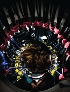

Image FROM “BIOIMAGING: WATCHING THE BRAIN AT WORK,” BY ROBERT J. COOPER, IN NATURE PHOTONICS, VOL. 8; JUNE 2014
Step aside, huge magnets and radioactive tracers—soon some brain activity will be revealed by simply training dozens of red lights on the scalp. A new study in Nature Photonics finds this optical technique can replicate functional MRI experiments, and it is more comfortable, more portable and less expensive.
The method is an enhancement of diffuse optical tomography (DOT), in which a device shines tiny points of red light at a subject's scalp and analyzes the light that bounces back. The red light reflects off red hemoglobin in the blood but does not interact as much with tissues of other colors, which allows researchers to recover an fMRI-like image of changing blood flow in the brain at work. For years researchers attempting to use DOT have been limited by the difficulty of packing many heavy light sources and detectors into the small area around the head. They also needed better techniques for analyzing the flood of data that the detectors collected.
Now researchers at Washington University in St. Louis and the University of Birmingham in England report they have solved those problems and made the first high-density DOT (HD-DOT) brain scans.
Via iPamba